Confocal laser scanning and spinning-disk confocal microscopy allow researchers to generate 3D images of organelles within living cells and examine changes that occur in cells over time. Used to image live cells and observe cell activity in real time.

Instruments Of Microscopy Microbiology
B View cell contents.

. Compound microscopes can see the nuclei of cells. Which microscope can be used to view live cells. For optical sectioning and Z-stacks the Apotome slider module transforms Cell Observer into a 3D workstation.
Used to study cancer cells artery plaques and biofilms. Scanning electron microscopy O phase-contrast microscopy O bright-field microscopy transmission electron microscopy. The image seen with this type of microscope is two dimensional.
Tissue and cells are typically placed in a petri dish on the microscope stage. The long working distance allows room for the petri dish since the objective lenses are beneath the stage rather than above it on an upright biological microscope. Electron microscopes can also magnify samples up to two million times while light microscopes are limited to 2000 times.
D all of these. Compound microscopes are light illuminated. A beam of electrons scans back and forth over the surface of a specimen coated with a thin film of metal.
These companies also offer customer-friendly technical support. However it has a low resolution. To help our students we have arranged bespoke microscopy packages with several reputable microscope suppliers worldwide.
You can view individual cells even living ones. Uses 2 different dyes. Based on structured illumination microscopy SIM Apotome generates extremely high-resolution optical sections of your live cells.
C The light gets reflected from the sides of the specimen and appears bright in dark background. A dissection microscope is light illuminated. 31 Related Question Answers Found What is the smallest thing we can see with an electron microscope.
The type of microscopy which can be used for visualizing live cells is phase contrast microscopy View the full answer Transcribed image text. As those beams move electrons are released from the specimen then ____ back to the viewing chamber. This microscope is the most commonly used.
B The stop disc prevents the entry of light from the central field and object is illuminated with beam of light. Uses two photons to illuminate a specimen. B Closest to the specimen.
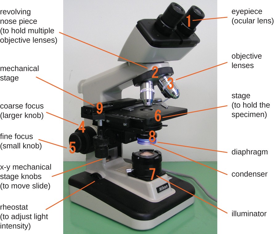
The objective lenses are the ones. A stereo microscope is used to. The Live Cell Imaging Microscope Most modern widefield epifluorescence spinning disk confocal or TIRF microscope setups rely on a similar set of optical and mechanical components and all imaging modalities are often used for live cell imaging.
E a scanning electron microscope because it can be used to observe whole cells without slicing them. C At the base of the microscope 9. A _____ electron microscope is used to view surface details of cells.
Light microscopes let us look at objects as long as a millimetre 10-3 m and as small as 02 micrometres 02 thousands of a millimetre or 2 x 10⁻⁷ m whereas the most. In effect a 3D image is formed. However biologists have been unable to unleash the high power of electron microscopes on living specimens.
D Observe cell cycle 8. Interprets the action of a sound wave sent through a specimen. Tissue culture microscopes are used to view live cells and tissue.
A Closest to the eye. It has high magnification. These longer working distances allow the ability to focus on the live cells through the bottom of the.
Typically magnification of scanning electron microscope is around 20000 times. This is used to study the behavior of living cells observe the nuclear and cytoplasmic changes taking place during mitosis and the. Confocal laser scanning microscopes only allow for a relatively slow image acquisition speed 3.
C Observe live cells. More powerful instruments such as an electron microscope can reveal the smallest components of organelles and even the. Some live cells are placed in a petri dish while others are grown in similar devices whatever the cell container being utilized live cell microscopes provide ample space for these live cells to grow.
A Adding disc called stop to the condenser will make bright field to dark filed. All our recommended professional microscope packages are designed to be suitable for Live Dry Blood Analysis and represent excellent quality at great prices. A Observe the surface of an object.
QUESTION 9 Which of the following types of microscopy can be used with live cells. Live cell microscopes also use objective lenses that offer longer working distances.

3 2a Microscopy Biology Libretexts
3 1 How Cells Are Studied Concepts Of Biology 1st Canadian Edition

0 Comments